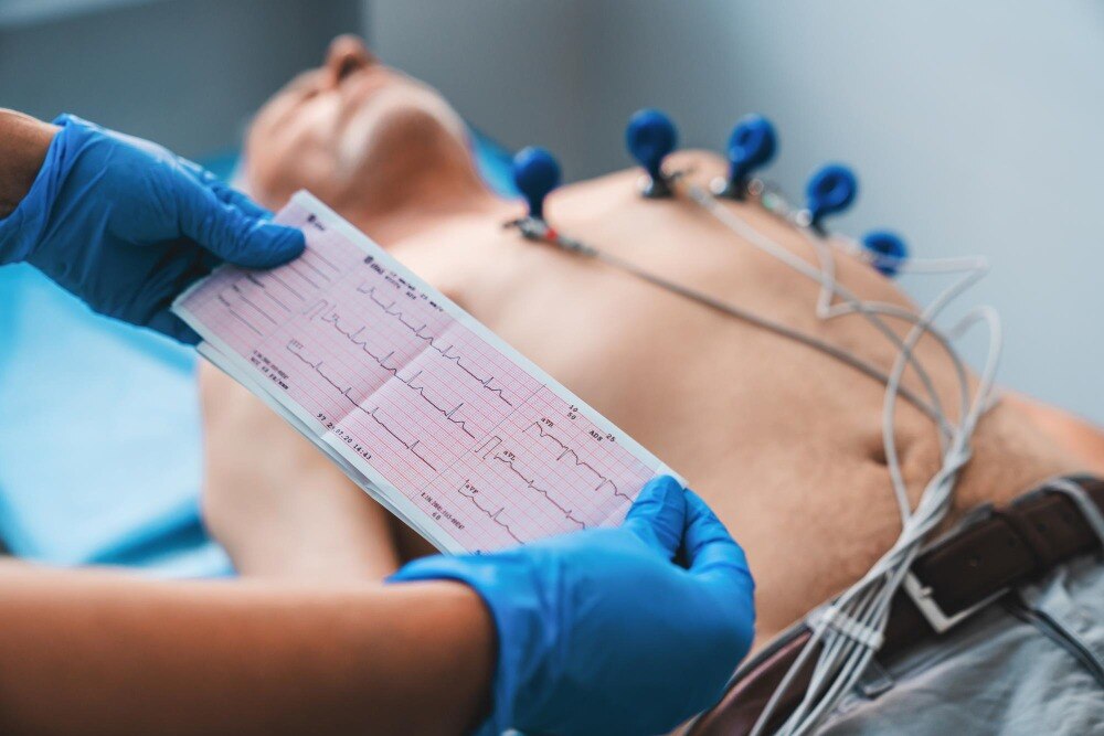
A brief ECG can show whether atrial and ventricular activity are timed normally, whether electrical impulses are delayed or blocked, and whether the tracing contains clues to ischemia, infarction, medication effects, or electrolyte disturbance. You will learn how the test is recorded, what its waveforms mean, why a normal result can miss intermittent symptoms, and when another form of monitoring or cardiac testing is needed.
Key takeaways
- A 12-lead ECG uses 10 electrodes to record the heart from 12 electrical views.
- A normal resting ECG cannot rule out an arrhythmia that occurs intermittently.
- Repeat the ECG during symptoms or use longer monitoring when episodes come and go.
- Clinicians confirm abnormal findings using symptoms, prior ECGs, examination and targeted tests.
What a 12-lead ECG records and how the test is performed
A 12-lead ECG records the heart’s electrical activity from 12 views using 10 physical electrodes: four on the limbs and six across the chest. It is not a picture of the heart, but it shows how electrical impulses start, travel, and recover.
- The P wave shows atrial activation.
- The PR interval measures conduction from the atria through the atrioventricular node to the ventricles.
- The QRS complex shows ventricular activation; its duration indicates how quickly that impulse spreads.
- The T wave shows ventricular recovery.
- The QT interval covers ventricular activation and recovery together and changes with heart rate.
The relationship between P waves and QRS complexes helps clinicians assess whether atrial impulses reach the ventricles regularly. The tracing can therefore reveal a fast or slow rate, an irregular rhythm, or delayed conduction, although the cause requires clinical interpretation.
For recording, you lie still on your back while the technician cleans and dries the skin, attaches electrodes to both limbs and six chest positions labelled V1 to V6, and connects lead wires. Remove clothing from your upper body, avoid lotion at electrode sites, and tell the technician about implanted devices and medicines.
The stickers may tug slightly when removed, but the test is painless and does not send electricity into your body.
A standard recording takes about 10 seconds. Keep still, relax your arms, and follow instructions about breathing; movement or shivering can create artefact. Correct electrode placement matters because misplaced V1 or V2 can produce a false pattern.
Which rhythm, conduction and activity changes can appear on an ECG?
An irregular pulse does not identify its cause. A 12-lead tracing must be examined for P waves, QRS timing, ventricular rate and the relationship between atrial and ventricular activity.
| ECG finding | What it can show | What it does not prove |
|---|---|---|
| Atrial rhythm | Atrial fibrillation, atrial flutter, premature beats and other ectopic rhythms | An irregular appearance alone does not confirm atrial fibrillation; artifact can mimic it |
| Ventricular rate | Bradycardia, tachycardia and some ventricular arrhythmias | A brief normal rate does not exclude episodes occurring later |
| Conduction | First-, second- or third-degree heart block, bundle-branch block and paced rhythm | An old conduction abnormality may be less urgent than a new change |
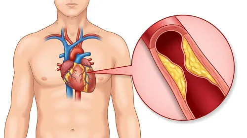
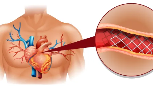
| Electrical activity | ST-segment or T-wave changes, infarction patterns, ventricular hypertrophy and QT prolongation | It cannot by itself establish a coronary blockage; potassium disturbances and medicines can produce similar changes |
A normal tracing shows no detectable abnormality during those recorded seconds, but it cannot rule out intermittent symptoms. An abnormal tracing needs comparison with earlier ECGs: a new modest change can matter more than a longstanding striking one.
Movement, muscle tension, loose electrodes and poor skin contact can create artifact that resembles arrhythmia or ST abnormality; inconsistency between leads or disappearance after electrodes are reapplied supports artifact.
A single-lead consumer recording can document rhythm during symptoms, but it cannot assess conduction, ischemia or infarction as comprehensively as a 12-lead ECG, and automated alerts require clinical confirmation.
Why a normal resting ECG can miss an intermittent arrhythmia
A normal office ECG can miss an arrhythmia because it usually records only about 10 seconds. If your heartbeat is regular during that snapshot, the tracing cannot show an episode that happened earlier, ended before recording, or starts hours later. Palpitations can therefore continue despite a normal result.
The monitor should match how often symptoms occur:
| Symptom interval | Monitor | How it helps |
|---|---|---|
| Several times daily or most days | Holter monitor | Records continuously, commonly for 24–48 hours, so it can match symptoms with the rhythm at that moment. |
| Weekly or less predictable episodes | Event monitor | Records when you activate it during symptoms; some models detect selected rhythms automatically over several weeks. |
| Daily to several-week episodes | Patch monitor | A small adhesive device records continuously for days or weeks without the wires of a standard Holter monitor. |
| Brief, occasional symptoms | Wearable ECG | Lets you capture a rhythm strip during an episode, but it may not provide continuous surveillance or a diagnostic-quality 12-lead tracing. |
| Rare fainting or palpitations over months | Implantable loop recorder | Sits beneath the skin and monitors for much longer, storing detected or patient-activated events. |
Tell the clinician how often symptoms occur, how long they last, and whether dizziness, fainting, chest pain, or breathlessness accompanies them. Seek urgent care for palpitations with fainting, severe breathlessness, or chest pressure; monitoring should not delay emergency assessment.
When an ECG needs to be repeated or paired with another test
A normal resting tracing does not rule out a problem outside the heart’s electrical system. Compare it with another test when symptoms persist, the ECG changes, or you need information about blood flow, pumping strength, valves, oxygen delivery, or heart-muscle injury.
| Test | What it adds | When it applies |
|---|---|---|
| Resting ECG | Rhythm, conduction, and electrical signs of ischemia or myocardial infarction | Repeat it when symptoms change, an unexpected result appears, or a prior tracing is unavailable |
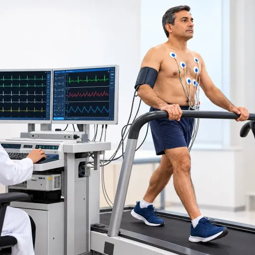
| Exercise stress test | Shows how rhythm and ST segments respond to exertion | Use for stable exertional chest pain after acute coronary syndrome has been excluded; do not use it to assess an emergency |
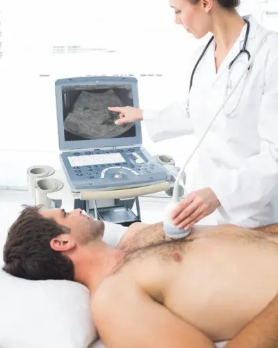
| Echocardiogram | Measures pumping function, chamber size, valve movement, and wall-motion abnormalities | Add it when breathlessness, a murmur, heart failure, or a new ECG abnormality needs structural assessment |
| Blood tests | Troponin detects heart-muscle injury; electrolytes and thyroid tests can reveal reversible rhythm triggers | Order them for suspected acute coronary syndrome, myocardial infarction, or unexplained arrhythmia |
| Cardiac imaging | Coronary CT angiography shows coronary narrowing; cardiac MRI can assess scar, inflammation, or tissue damage | Choose it when coronary anatomy or myocardial tissue cannot be judged from ECG and echocardiogram |
A nondiagnostic first ECG does not end an acute coronary syndrome assessment. Repeat ECGs promptly when suspicion remains or pain changes, because ischemic abnormalities can evolve; compare each tracing with older records. A new change matters more than a longstanding bundle-branch block or paced pattern. Verify electrode placement before accepting an unexpected result.
How clinicians confirm an ECG finding and decide what happens next
A new ECG change is more concerning when it matches current symptoms, such as chest pressure, fainting, severe breathlessness or sustained palpitations. If suspected acute coronary syndrome remains possible, clinicians repeat the tracing promptly, especially after symptoms change, because ischemic abnormalities can evolve.
Clinicians check:
- Prior ECG comparison: an old bundle-branch block, paced rhythm or persistent ST-T pattern may be less urgent than a new, even modest, change.
- Electrode placement: V1 and V2 placed too high can mimic poor R-wave progression or a septal infarction. Limb-lead reversal can imitate axis or ischemic changes, so staff repeat an unexpected tracing after verifying every lead.
- Implanted devices: a pacemaker ECG has pacing spikes and altered QRS patterns that require device history and, when needed, formal pacemaker interrogation rather than comparison with an untreated rhythm.
- Medications and blood results: potassium changes and another electrolyte disturbance can alter T waves, conduction and QRS width. Medicines can cause QT prolongation, so clinicians assess the QTc, heart rate and correction method instead of using the raw QT alone.
- Specialist review: an automated alert or single-lead recording can document a rhythm during symptoms, but a clinician must confirm it with a diagnostic 12-lead ECG and the full clinical context.
For an unexplained new finding, medication-related QTc concern or pacemaker ECG, a cardiologist such as Dr Vijay Bang Cardiologist in Mumbai can integrate the tracing with symptoms, records, laboratory results and device information before recommending monitoring, treatment or urgent assessment.
Related service
ECG (Electrocardiogram) An ECG (Electrocardiogram) is a simple, painless test that records the electrical activity of the heart. View service → |
Frequently asked questions
What does a 12-lead ECG record?
It records the heart’s electrical activity from 12 views using 10 physical electrodes: four on the limbs and six on the chest. The tracing shows how impulses start, travel through the heart, and recover.
Which changes can an ECG show?
An ECG can show abnormal rhythms, slow or fast rates, conduction delays or blocks, and changes in electrical activity linked with conditions such as reduced blood flow or previous heart damage.
Why can a normal resting ECG miss an arrhythmia?
A resting ECG captures only a short period. If the abnormal rhythm is not occurring during the recording, the tracing can look normal even when symptoms are caused by an intermittent arrhythmia.
When is another test needed after an ECG?
Clinicians may repeat the ECG during symptoms or pair it with ambulatory monitoring, such as a Holter monitor or event recorder, when episodes are intermittent. They use other tests when the ECG suggests a structural or circulation problem.