A 2D echo combines ultrasound images with Doppler and other measurements to show how the heart is built, how its chambers and valves work, and how blood moves through it. By the end, you will know what happens during the test, what to bring, which report terms matter, and when a finding needs further clinical evaluation.
Key takeaways
- A 2D echo shows moving heart anatomy without ionising radiation.
- M-mode measures movement; Doppler evaluates blood flow through chambers and valves.
- Wear clothing that allows easy access to your chest during the scan.
- Interpret ejection fraction alongside valves, chambers, wall motion, and symptoms.
What a 2D Echo Shows Inside the Heart
A routine 2D echo test uses reflected sound waves to create moving, cross-sectional images of the heart without ionising radiation. During a transthoracic echocardiogram, an ultrasound transducer sends and receives these echoes through the chest. The examination also commonly includes M-mode and Doppler imaging, so it evaluates movement and blood flow as well as anatomy.
It can assess:
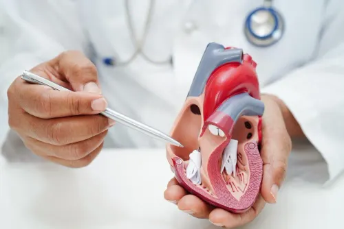
- The four chambers, including their size, shape and overall pumping movement.
- The septum and ventricular walls, including areas that move abnormally after heart muscle damage.
- The mitral, tricuspid, aortic and pulmonary valves, showing how they open, close and allow or leak blood.
- The pericardium, the thin sac around the heart, including fluid collecting around it.
- The aortic root and, when visible, the inferior vena cava, which can help assess nearby anatomy and right-sided filling conditions.
Doppler measurements show the direction and speed of blood moving through chambers and valves. They help estimate pressure differences, assess valve narrowing or regurgitation, and describe how the left ventricle fills between beats.
A high ejection fraction alone does not prove normal heart function: a resting scan can still show abnormal relaxation, valve disease or reduced right-ventricular performance.
A resting scan can reveal the effect of coronary disease through a regional wall-motion abnormality, but it cannot directly show a narrowed coronary artery. When heart borders are difficult to see, an ultrasound-enhancing agent can improve the image without turning the examination into a CT or MRI scan.
How the Scan Measures Pumping, Valves and Blood Flow
Ejection fraction shows how much blood the left ventricle ejects with each contraction: an EF of 60% means 60% of the blood present before the beat leaves during that beat. It is a pumping measure, not a complete score of heart health.
A preserved EF can coexist with heart failure, abnormal relaxation, significant valve disease, or weak right-ventricular function.
| Measurement | What it shows | What an abnormal result may suggest |
|---|---|---|
| Ejection fraction | Left-ventricular squeeze | Reduced pumping function |
| Wall-motion abnormality | Whether each ventricular segment contracts evenly | Previous or active coronary-related injury; a resting echo does not prove a coronary narrowing |
| Chamber size and wall thickness | The heart’s shape and workload | Enlargement, pressure overload, or remodelling |
| Filling pattern | How the ventricle relaxes and receives blood | Impaired relaxation or raised filling pressures |
| Heart valve opening | How freely a valve allows blood to pass | Narrowing, called stenosis |
| Valve regurgitation | Whether blood leaks backward through a valve | Leakage that can enlarge or strain a chamber |
Doppler adds the blood-flow evidence that moving pictures alone cannot provide. It measures flow direction and velocity across valves and through chambers; a high-velocity jet can estimate a pressure difference using approximately 4 × velocity², but velocity alone cannot grade stenosis or regurgitation reliably.
The report may also include right-sided pressure estimates and observations of blood returning through the inferior vena cava. Blood pressure, hydration, heart rate, rhythm, loading conditions, and image quality affect these values, so a small change between scans does not automatically mean the heart has improved or deteriorated.
What Happens During the Appointment
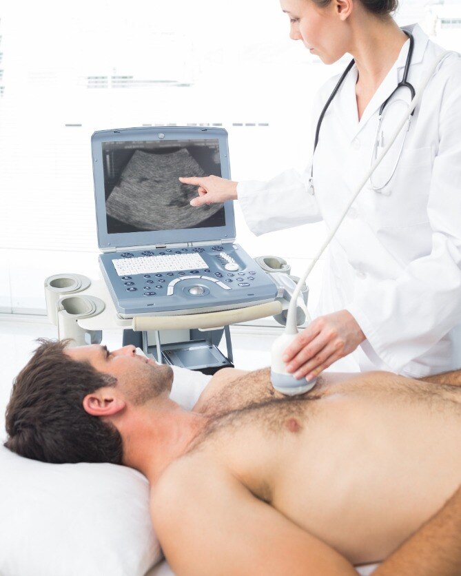
You remain awake throughout a routine 2D echo procedure. After you remove clothing from the upper body, you lie in a left-side position on the examination bed. The sonographer applies warm gel and moves a handheld transducer across your chest to capture images through several acoustic windows.
You may be asked to hold your breath briefly or shift position so the heart is easier to see.
- You may feel firm pressure as the transducer is moved between the ribs; the gel feels wet but is wiped off afterward.
- The machine produces moving images and records Doppler measurements of blood flow. You may hear pulsing or whooshing sounds, but the scan does not use radiation.
- A routine appointment usually takes about 30 to 60 minutes. Difficult images, an irregular rhythm, or extra measurements can extend the session.
You usually need no special preparation. No fasting for 2D echo is required, so you can normally eat, drink and take prescribed medicines unless the testing centre gives different instructions. Wear clothing that allows easy access to your chest, and bring previous heart-test reports if requested.
Do not assume every heart ultrasound follows these instructions. A transesophageal echocardiogram places the probe in the oesophagus and usually requires fasting and sedation, so follow its separate appointment instructions.
How to Prepare and Help the Sonographer Get Useful Images
Give the imaging team your main symptoms, heart diagnosis, previous surgery, and the reason for the scan. Bring the prior echo report and medication list if available; earlier measurements help the cardiologist compare chamber size, valve findings, and pumping function rather than interpret one examination in isolation.
- Tell the sonographer about an implanted device, including a pacemaker, defibrillator, valve replacement, or repair. The device and its position can affect which views are useful.
- Explain if you have emphysema, other lung disease, obesity, chest-wall changes, or difficulty lying on your left side. These details help the operator plan alternative windows.
- Report chest pain, breathlessness, fainting, palpitations, or a recent change in exercise tolerance. Symptoms give the images clinical context.
- Follow the appointment instructions about clothing and medicines. A routine transthoracic echo usually needs no fasting, but ask before eating or taking medicine if the appointment might change to a transesophageal echocardiogram.
- Hold your breath or change position when asked. A few seconds of stillness can separate a clear valve edge from a blurred one.
Poor acoustic windows can produce limited echocardiogram views, leaving a chamber, valve, or wall-motion segment difficult to assess. An unseen structure is not automatically normal. Image quality also affects measurements such as ejection fraction; when borders are clear, biplane Simpson’s method is more dependable than a quick visual estimate.
A transesophageal study places the probe in the esophagus and uses fasting and sedation to answer questions that chest views cannot.
How to Read the Report Without Overinterpreting One Number
A 55% left-ventricular ejection fraction does not certify that the heart is healthy. It measures the percentage of blood pushed out of the left ventricle with each beat, while valve disease, abnormal relaxation, heart-failure with preserved EF, or weak right-ventricular function can still cause symptoms.
1. Check how the measurement was obtained. Biplane Simpson’s method traces the left-ventricular lining in two views and is more reproducible than a visual estimate when images are clear. Poor windows, irregular rhythms, and beat-to-beat variation reduce confidence, so compare the number with earlier studies rather than treating a small change as deterioration.
2. Read valve findings as a group. A gradient, velocity, or colour-Doppler jet alone cannot grade valve disease; anatomy, valve area, chamber size, and the ventricle’s response also matter. Ask what the combined assessment means if one value appears alarming.
3. Treat a pulmonary artery pressure estimate as an estimate. It usually combines the tricuspid-regurgitation jet with an inferior-vena-cava assessment, and a weak signal or difficult right-sided measurements can make it substantially inaccurate.
4. Look beyond EF for diastolic function and right-ventricular performance. Mitral inflow, tissue-Doppler e′, E/e′, left-atrial volume, TAPSE, S′, and visual assessment work together; atrial fibrillation, fast heart rate, pacing, or poor signals can make conclusions uncertain.
A pericardial effusion, new wall-motion abnormality, rising pressure estimate, or symptoms such as breathlessness and fainting deserves prompt discussion with a cardiologist. Dr Vijay Bang Cardiologist in Mumbai can help interpret the complete report alongside your symptoms, examination, ECG, and prior scans.
Frequently asked questions
What does a 2D echo show inside the heart?
It creates moving cross-sectional images of the heart, showing its chambers, walls, valves, and overall structure.
How does a 2D echo measure pumping and blood flow?
M-mode records heart movement, while Doppler imaging assesses the direction and speed of blood flow through the heart.
What happens during a 2D echo appointment?
During a transthoracic echocardiogram, a sonographer moves an ultrasound transducer across your chest to send and receive reflected sound waves.
How should you prepare for a 2D echo?
Follow the appointment instructions, wear clothing that permits chest access, and tell the sonographer if positioning causes discomfort.
How should you read a 2D echo report?
Read ejection fraction with findings about chamber size, wall motion, valves, and blood flow instead of interpreting one number alone.