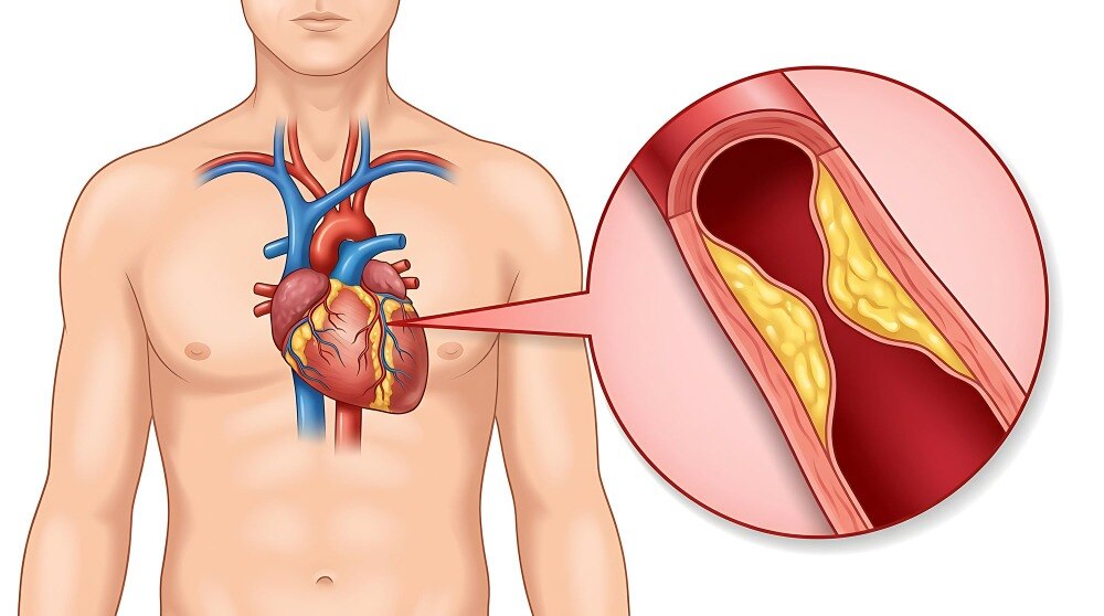
Angiography is not automatically the next step for every episode of chest pain or every abnormal test. By separating emergency symptoms from stable symptoms, understanding when non-invasive tests come first, and weighing anatomy against blood-flow evidence, you can understand why a cardiologist recommends it and what the result could change.
Key takeaways
- Seek emergency care for persistent chest pressure, breathlessness, sweating, fainting, or spreading pain.
- Use ECG, stress testing, or CT coronary angiography first when symptoms are stable.
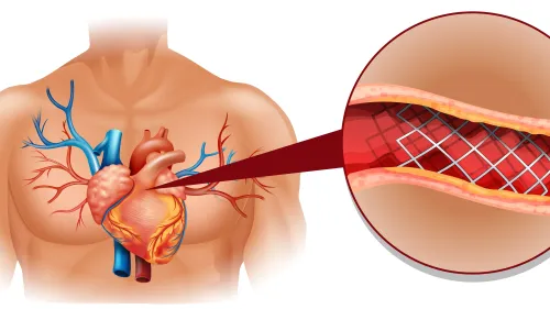
- An angiogram shows where coronary arteries narrow and whether blood flow is restricted.
- Ask about fasting, medicines, kidney tests, contrast risks, and recovery before the procedure.
Which symptoms require emergency angiography rather than a routine appointment?
Persistent chest pressure, breathlessness, sweating, fainting, or pain spreading to your arm, back, shoulder, or jaw requires emergency care, especially if you have known heart disease. Do not wait for a routine appointment.
| Emergency angiography | Urgent coronary angiography | Planned outpatient assessment |
|---|---|---|
| STEMI with suspected artery occlusion; performed immediately as part of primary PCI | NSTE-ACS with very high-risk symptoms and signs; performed promptly during the admission | Stable symptoms that settle with rest and have no high-risk features |
| No treadmill test or coronary CT scan first | No outpatient testing delay when instability or ongoing ischaemia is present | ECG, echocardiography, stress testing, or coronary CT angiography may guide the decision |
With STEMI, emergency angiography usually forms part of primary PCI to reopen the culprit artery. Do not wait for a treadmill test or coronary CT scan.
NSTE-ACS symptoms and signs that prompt urgent coronary angiography include:
- Shock or haemodynamic instability
- Recurrent or refractory chest pain
- Life-threatening arrhythmia or cardiac arrest
- Acute heart failure caused by ongoing ischaemia
- Mechanical complications after myocardial infarction
A first normal ECG or one negative high-sensitivity troponin does not safely exclude acute coronary syndrome. Clinicians may repeat the ECG and use serial ECG and high-sensitivity troponin testing at timed intervals.
If symptoms remain stable, settle with rest, and lack high-risk features, planned outpatient assessment is usually appropriate. A cardiologist then matches your symptoms, examination, ECG, and test results to the safest next step.
When should non-invasive testing come before an angiogram?
In stable chest pain, invasive angiography is not usually a screening test or the automatic first test because it requires arterial catheterisation, contrast, and radiation. Doctors begin with non-invasive cardiac tests to estimate risk and identify a safer, more useful next step.
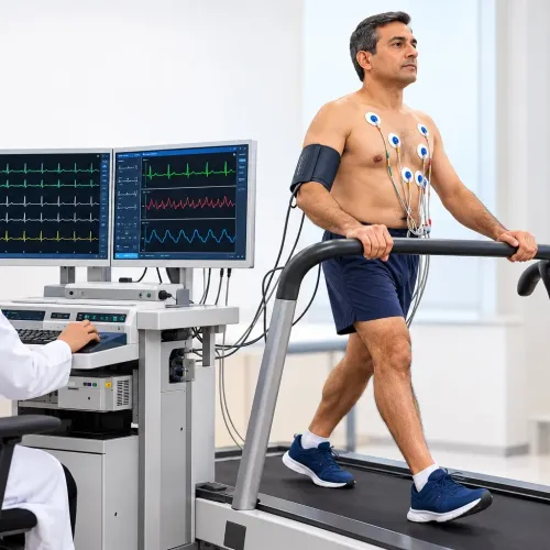
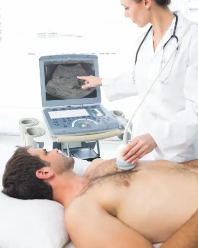
1. An ECG can show prior infarction, ischaemia, or rhythm problems. An echocardiogram assesses left-ventricular function, valve disease, and other causes of pain or breathlessness. A treadmill stress test examines ECG changes and symptoms during exertion; stress echocardiography adds exercise or medication-induced wall-motion changes, helping assess whether exertion produces ischaemia.
2. Coronary CT angiography, or CCTA, shows coronary anatomy, plaque, and narrowing. It is useful for excluding obstructive disease, while functional tests assess whether a blockage limits blood supply during exertion.
CCTA becomes less reliable with heavy calcification, a fast or irregular rhythm, inability to hold your breath, severe renal dysfunction, or stents and bypass grafts that are difficult to interpret.
3. The threshold for invasive angiography rises or falls with your age, symptoms, cardiovascular risk factors, prior coronary disease, ECG findings, left-ventricular function, symptom severity, and the quality of earlier test results. A vague, stable symptom with a good-quality reassuring test rarely warrants catheterisation.
4. Invasive testing becomes more reasonable with very high clinical likelihood, angina at low exertion, severe or refractory symptoms despite treatment, or a non-invasive result that would change treatment. That is when angiography is recommended for stable chest pain rather than used routinely.
What can an angiogram prove about a suspected blockage?
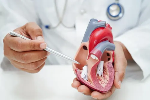
An invasive angiogram shows the shape and diameter of the coronary artery lumen. It shows anatomical narrowing versus blood-flow restriction, but it does not directly measure blood flow to heart muscle or prove that every visible narrowing causes ischaemia.
Visual estimates become unreliable when the artery has:
- An intermediate coronary stenosis
- Diffuse disease rather than one clear lesion
- Serial stenoses that affect pressure together
- Left-main disease
- Vessel overlap or foreshortening that hides the true lesion length
When severity remains uncertain, FFR or iFR measures the pressure effect of a narrowing and helps determine whether it is functionally important. IVUS or OCT provides cross-sectional detail about the vessel wall, plaque, calcium, and lumen, which can clarify anatomy and guide complex PCI.
An abnormal stress test does not automatically mean that you need a stent. Doctors combine symptoms, anatomy, objective ischaemia, pressure measurements, ventricular function, and procedural risk before recommending revascularisation.
A normal or non-obstructive angiogram also does not make persistent chest pain imaginary. INOCA can involve coronary microvascular dysfunction, including microvascular angina, or artery spasm causing vasospastic angina. Selected patients need coronary function testing to identify these problems; placing a stent in a vessel without a flow-limiting blockage will not treat them.
How do angiography findings change treatment decisions?
An angiogram is a diagnostic test, not an automatic angioplasty or stent appointment. Angiography results show coronary anatomy, but treatment also depends on symptoms, objective ischaemia, prognosis, guideline-directed medical therapy, ventricular function, and procedural risks.
- Medicines and risk-factor control: Use this when disease does not justify revascularisation. Treatment can include cholesterol-lowering therapy, blood-pressure control, smoking cessation, diabetes management, exercise, and anti-anginal medicines.
- PCI with a stent: Consider catheter treatment when symptoms remain limiting, the anatomy is suitable, and objective ischaemia supports treating the narrowing. A visible blockage alone does not prove that a stent will improve outcomes.
- Bypass surgery: Coronary artery bypass grafting may offer greater benefit when disease is extensive, involves the left main or several vessels, or coexists with reduced ventricular function or diabetes. Surgical risk, graft suitability, frailty, and the patient’s preferences also matter.
The main trade-off of invasive angiography is exposure to complications without a guaranteed need for treatment. Discuss these procedural risks before consent:
- Bleeding or bruising and artery damage at the catheter site
- Contrast-associated kidney injury or an allergic-like contrast reaction
- Radiation exposure and temporary or serious arrhythmia
- Stroke, heart attack, or rare death
Risk rises during emergency treatment and with kidney disease, older age, frailty, or serious coexisting illness. A normal or non-obstructive result still guides medicines and follow-up rather than ending the evaluation of persistent symptoms.
What should you arrange and ask before planned angiography?
Your angiography preparation starts with a complete medication review. Bring every prescription, over-the-counter medicine, supplement, dose, and timing. Discuss anticoagulants and antiplatelet medicines before the appointment; do not stop either without instructions. Ask exactly how to adjust diabetes medicines, complete kidney-function testing when requested, and follow the fasting instructions.
Report pregnancy or possible pregnancy, active bleeding, severe anaemia, advanced kidney disease, or a previous contrast allergy. These details can change the timing, medicines, contrast choice, or need for additional precautions.
The usual process involves local anaesthetic, catheter entry through the radial artery at the wrist or the femoral artery in the groin, contrast injection, X-ray imaging, and monitoring afterward. The procedure often takes less time than the observation period. Discharge timing depends on the access site, findings, and whether treatment occurs during the same admission.
Arrange transport home; possible overnight observation and temporary restrictions on lifting or driving may affect your plans.
Ask these consent questions before signing:
- Why is angiography recommended for me?
- What alternatives exist, including medical treatment or non-invasive testing?
- Could same-session PCI, such as balloon treatment or stenting, happen if a blockage is found?
- Who will review the findings with me and explain the follow-up plan?
A cardiology service such as Dr Vijay Bang Cardiologist in Mumbai can help connect the recommendation, preparation, findings, and follow-up, but the need for angiography depends on your individual clinical assessment.
Related service
Coronary Angiography Doing a good angiography is also not an easy task. People search for the best angiography doctors in Mumbai or in India and that is when they call Dr... View service → |
Frequently asked questions
Which symptoms require emergency angiography rather than a routine appointment?
Persistent chest pressure, breathlessness, sweating, fainting, or pain spreading to the arm, back, shoulder, or jaw requires emergency care. The emergency team decides whether urgent angiography is needed after an ECG, blood tests, and clinical assessment.
When should non-invasive testing come before an angiogram?
When symptoms are stable and there is no evidence of an acute heart attack, doctors may begin with an ECG, echocardiogram, stress test, or CT coronary angiography. The choice depends on your symptoms, risk factors, kidney function, and previous test results.
What can an angiogram prove about a suspected blockage?
A coronary angiogram shows the location and severity of narrowing in the coronary arteries, identifies fully blocked vessels, and helps assess blood flow. It does not by itself explain every type of chest pain.
How do angiography findings change treatment decisions?
Mild or non-obstructive narrowing may lead to medicines and risk-factor control. A significant, suitable narrowing may support angioplasty and stent placement, while complex or extensive disease may require discussion of bypass surgery.
What should you arrange and ask before planned angiography?
Ask whether to fast, which medicines to take or stop, whether kidney-function testing is needed, how contrast allergy risk will be managed, and when you can drive, work, or resume exercise.