A TMT shows how your heart’s electrical activity, rhythm, blood pressure, symptoms, and exercise capacity respond as physical effort increases. You will learn what happens on the treadmill, how to prepare, what the results can and cannot prove, and when another cardiac test is more suitable.
Key takeaways
- Wear comfortable clothes and shoes; avoid a heavy meal before the test.
- TMT tracks ECG changes, blood pressure, rhythm, symptoms, and exercise capacity.
- Tell the testing team about chest pain, breathlessness, dizziness, or medication use.
- Another test may be better when exercise is unsafe or the ECG is difficult to interpret.
What does a TMT measure during exercise?
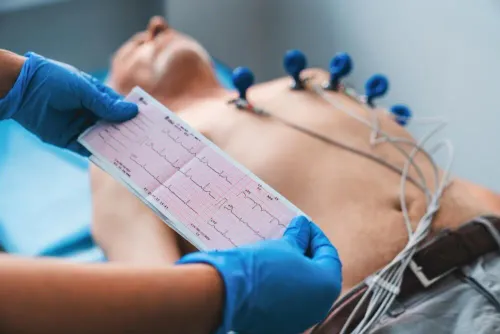
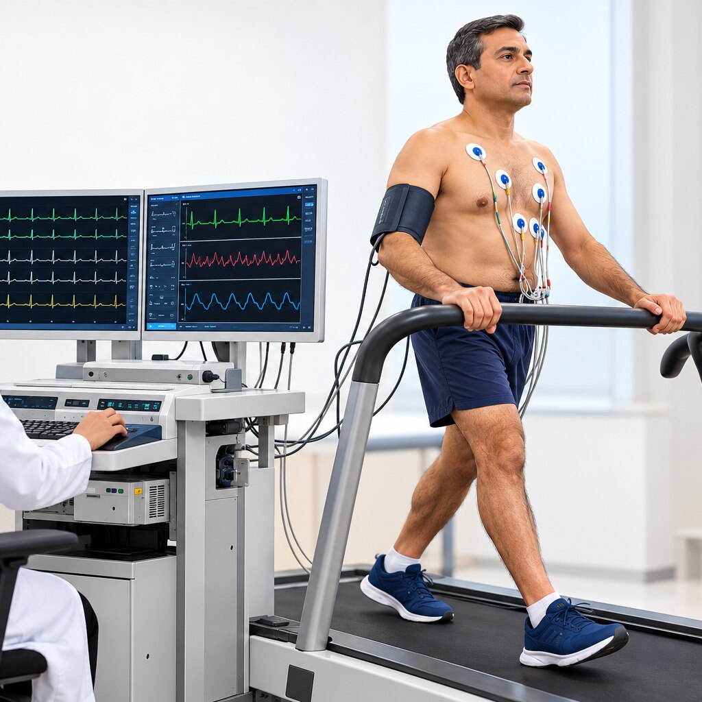
A TMT test shows how the heart responds when physical demand rises. It begins with a resting 12-lead ECG, then tracks the exercise ECG, heart rate, blood pressure, rhythm, symptoms, and exercise capacity as you walk or pedal under progressively heavier workloads.
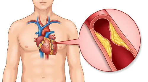
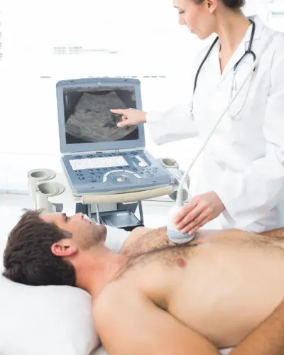
This makes it useful for assessing heart function during physical activity, especially whether exertion brings chest discomfort, breathlessness, abnormal rhythms, an inadequate heart-rate response, or ECG changes that suggest reduced blood flow. It does not directly show the heart’s pumping anatomy or prove how well each chamber and valve works.
| Question | Best-suited test | What it shows |
|---|---|---|
| Does exertion trigger electrical signs of reduced blood flow? | TMT or exercise ECG | ECG changes, symptoms, heart-rate and blood-pressure responses |
| How much blood does the left ventricle pump? | Echocardiogram | Ejection fraction, chamber size, wall motion, and pumping function |
| Are valves, chambers, or surrounding structures abnormal? | Echocardiogram or cardiac imaging | Valve movement, chamber anatomy, wall motion, and structural disease |
Treadmill walking also cannot reproduce every trigger. Cycling, lifting, hills, heat, or a job-specific effort can produce different demands; if symptoms occur only during one activity, reproducing that workload or choosing stress imaging may provide a clearer answer.
Certain baseline ECG patterns, including left bundle-branch block or ventricular pacing, can also make exercise ECG interpretation unreliable.
What happens as the treadmill workload increases?
The workload rises in measured stages, not in one sudden burst. After you remove clothing from the chest, a technician places adhesive electrodes for a 12-lead ECG, checks the signal, and records your resting heart rate, rhythm, blood pressure, and symptoms while you stand still.
You then walk while the ECG records continuously and blood pressure is checked at intervals. The Bruce protocol increases speed and incline every three minutes; the modified Bruce protocol begins with gentler stages for people with low fitness, limited mobility, or reduced exercise tolerance.
Tell the technician immediately about chest discomfort, breathlessness, dizziness, leg pain, or unusual fatigue. Exercise capacity is recorded in METs: 1 MET represents resting oxygen use, about 3.5 mL/kg/min.
The test ends when you reach the planned target heart rate or when safety requires an earlier stop:
- Chest pain or severe breathlessness
- Dizziness, faintness, or marked exhaustion
- Concerning ECG changes or a dangerous rhythm
- An abnormal blood-pressure response
- Inability to maintain a safe walking pace
You then stand or sit while ECG, heart rate, blood pressure, and symptoms continue under observation. Recovery matters: delayed heart-rate reduction, persistent symptoms, rhythm changes, or ongoing ECG abnormalities can add important information even after the treadmill stops.
How should you prepare for the appointment?
Plan 30–60 minutes for the appointment. The exercise portion is brief but progressively demanding: you will walk faster and possibly uphill while reporting chest discomfort, breathlessness, dizziness, or unusual fatigue. Wear comfortable clothing and walking shoes; avoid applying lotion or oil to your chest because it can loosen the ECG electrodes.
- Eat a light meal 2–3 hours beforehand, unless the clinic gives different instructions. Avoid a heavy meal immediately before testing.
- Do not smoke on the morning of the test, particularly in the hours beforehand. Nicotine can raise heart rate and blood pressure.
- Ask whether to avoid coffee, tea, energy drinks, or other caffeine before arrival; follow the testing centre’s specific rule.
- If you have diabetes, confirm instructions for insulin, sulfonylureas, and other diabetes medicines before fasting or delaying food. Bring your glucose meter and a fast-acting carbohydrate if the clinic advises it.
- Take prescribed medicines only as instructed. Beta blockers and other rate-limiting drugs can blunt the heart-rate response, but never stop them independently; the prescribing clinician decides whether withholding a dose serves the test’s purpose.
Call before attending if you have new or ongoing chest pain, unstable angina, fainting, severe breathlessness, an uncontrolled symptomatic rhythm, decompensated heart failure, or a recent acute illness. A clinician must postpone testing for conditions such as acute myocardial infarction, pulmonary embolism, myocarditis, pericarditis, severe symptomatic aortic stenosis, or aortic dissection.
How are TMT findings interpreted?
TMT findings are interpreted as a pattern, not a pass or fail. An abnormal symptom, ECG change, or blood-pressure response means more in someone with typical exertional chest pain than in someone with low pretest likelihood. A positive TMT does not prove a blocked coronary artery; false-positive and false-negative results occur.
Interpretation considers:
- Symptoms: chest pressure, breathlessness, dizziness, or leg fatigue; when they began; and whether they limited exercise.
- Exercise duration and METs: these show achieved capacity, while fitness, age, medicines, and mobility affect the result.
- Heart rate: a rising rate supports adequate effort; a blunted rise can reflect medicines, conduction disease, or limited effort.
- Blood-pressure response: pressure should rise with workload; a fall or inadequate rise warrants attention.
- Rhythm and recovery: new arrhythmias, persistent symptoms, delayed heart-rate or ECG recovery, and recovery blood pressure add context.
- ST-segment changes: horizontal ST depression or downsloping ST-segment depression, measured about 60–80 milliseconds after the J point, is more concerning when it is clear, symptom-linked, and present in relevant leads.
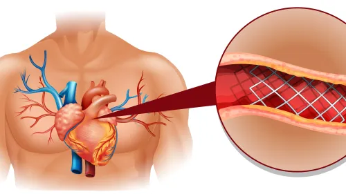
The Duke treadmill score combines exercise time, ST-segment deviation, and exercise angina to estimate risk; it does not show coronary anatomy. An abnormal or inconclusive result may require stress imaging, coronary CT angiography, or coronary angiography.
Dr Vijay Bang Cardiologist in Mumbai can weigh those findings against your symptoms, medicines, baseline ECG, and cardiovascular risk before selecting further evaluation.
Who benefits from a TMT, and when is another test better?
A TMT is most useful when a clinician has a defined question, such as:
- exertional chest discomfort
- unexplained breathlessness
- suspected exercise-induced arrhythmia
- a specific question about exercise capacity or response
Routine exercise-ECG screening is a poor choice for symptom-free, low-risk adults without known cardiovascular disease. False-positive results can lead to repeat tests or invasive procedures without providing a net benefit.
Reaching an age-predicted maximum heart rate does not prove that your heart is healthy. The estimate is imprecise, and the test may stop earlier because of symptoms, fatigue, an abnormal blood-pressure response, arrhythmia, or concerning ECG changes.
A treadmill walk may not reproduce symptoms triggered by cycling, lifting, hills, or heat. If one activity reliably causes symptoms, reproducing that workload—or choosing stress imaging—can provide more useful evidence.
A resting ECG can make exercise-ECG changes difficult to interpret when it shows:
- left bundle-branch block
- ventricular pacing
- substantial baseline ST depression
- left-ventricular hypertrophy with strain
In these situations, stress imaging or coronary CT angiography may be preferred. Stress imaging adds information about blood flow or heart-wall movement, while coronary CT angiography shows coronary anatomy.
An abnormal TMT does not prove that an artery is blocked. The result must be weighed against symptoms and pretest likelihood, because false-positive and false-negative results occur.
Frequently asked questions
What does a TMT measure during exercise?
A TMT measures the heart’s electrical activity, heart rate, blood pressure, rhythm, symptoms, and exercise capacity during increasing physical demand.
How should you prepare for a TMT appointment?
Wear comfortable walking shoes and loose clothing, avoid a heavy meal before testing, and ask whether you should adjust any medicines.
How are TMT findings interpreted?
A clinician compares the resting and exercise ECG with workload, heart-rate response, blood-pressure response, symptoms, rhythm, and recovery after exercise.
When is another test better than a TMT?
Another test may be more suitable when you cannot exercise safely, have an ECG that limits interpretation, or need anatomical or imaging information.